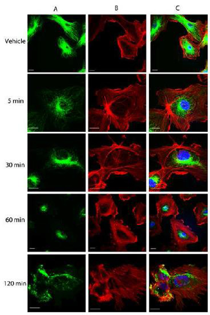
Figure 2.
Representative confocal images of the morphological effects of OXi8006 treatment on activated HUVECs. Monolayers of rapidly growing HUVECs on gelatin coated glass coverslips were treated with vehicle or 10 nM OXi8006 for the indicated times (5, 30, 60 and 120 min). Endothelial cells were fixed and stained with (A) anti-α-tubulin antibody (green, microtubules), (B) Texas red conjugated phalloidin (red,actin) and (C) DAPI (blue, nuclei), merged image. Bars, 20 µm. (For interpretation of the references to color in this figure legend, the reader is referred to the web version of this article.)

