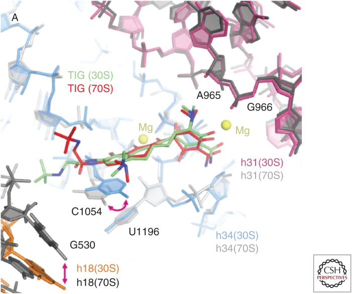
Figure 2.
Alternative binding modes of tigecycline at the primary ribosomal-binding site. Alternative tigecycline-binding modes in the 30S (green) and 70S (red) structures are shown, superimposed within the primary tetracycline-binding site. Key nucleotides (G530, A965, G966, C1054, U1196) and helices (h18, h31, h34) are shown in both structures. (From Schedlbauer et al. 2015; reprinted, with permission, from the American Society for Microbiology © 2015.)

