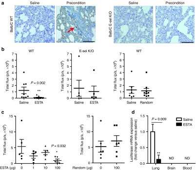
Figure 4.
ESTA inhibits the development of hematogenous metastasis. (a) The induction of E-selectin expression on the pulmonary capillary after preconditioning in Balb/C wild type mice (left), but not in E-selectin K/O mice (right). Scale bar represents 100 µm. (b) ESTA inhibits the development of metastasis in a syngeneic mouse model of breast cancer metastasis. Six-week-old female Balb/C or E-selectin K/O mice were preconditioned for a week with i.p. injections of conditioned media. The mice then received a single i.v. injection of ESTA (100 µg), random aptamer (100 µg), or saline, followed by 4T1-Luc cells (3 × 10⁴). Lung metastasis formation was measured by bioluminescent image 3 weeks after cancer cell inoculation. The data represent mean ± SEM. Statistical significance was determined by nonparametric Mann–Whitney test. (c) Effective dose of ESTA for the prevention of hematogenous metastasis. Six-week-old female athymic nu/nu mice (n = 5–6) was preconditioned with i.v. injection of VEGF. Two hours later, the mice were injected with either saline, various amounts of ESTA (1–100 ng), or random aptamer (100 ng), and then with MDA-MB-231-Luc (3 × 10⁴/100 µl saline). Bioluminescence was measured 3 weeks later. The data represent mean ± SEM. Statistical significance was determined by Mann–Whitney test. (d) Blockade of lung metastasis by ESTA does not cause a relocation of metastasis. Whole organs (lung and brain) and blood were collected 12 days after cancer cell injection. Luciferase mRNA was detected by 80 cycles of qRT-PCR and normalized by GAPDH. The data represent mean ± SD. Statistical significance was determined by Student's t-test.

