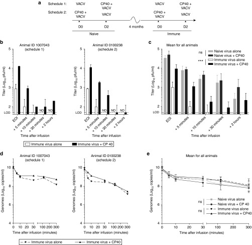
Figure 4.
Complement inhibition in a cynomolgus macaque model stabilizes vaccinia virus in the blood of immune animals. Vaccinia virus (1 × 10⁸ pfu) was infused using a central venous line over the course of 30 minutes. As per the treatment schedule in a, animals received a bolus intravenous dose of CP40 (2 mg/kg) immediately prior to virus treatment. Infectious virus in the blood was measured by plaque assay on samples taken at various time points after the end of the infusion (EOI). Blood titers for two representative immune animals are shown in b and are represented as technical replicate means ± SD. Mean titers for all naive and immune animals are shown in c and are represented as group means ± SD (n = 4 macaques). Genome content of blood collected at various time points after the EOI, as measured by qPCR using primers against the viral gene E3L. Genome concentrations in the blood are shown for two representative animals are shown in d and are represented as technical replicate means ± SD. Mean concentrations for all naive and immune animals in e and are represented as group means ± SD (n = 4 macaques). VACV, vaccinia virus, ND, not detected, LOD, limit of detection (***P < 0.001, ns = P > 0.05).

