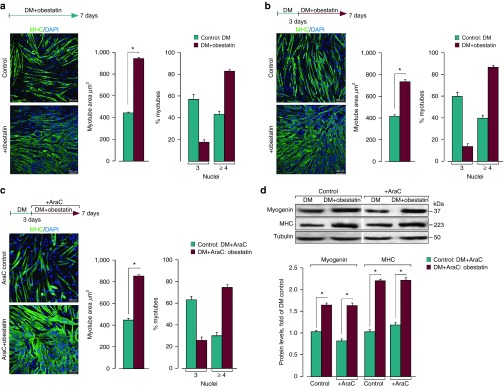
Figure 8.
The hypertrophic response to obestatin is not associated with the addition of new myonuclei via proliferation and further fusion. (a) Left panel, immunofluorescence detection of MHC and DAPI in C2C12 myotube cells under DM (control) or DM + obestatin (5 nmol/l) at the 7-day point after stimulation. Right panel, the myotube area (µm2) and the number of nuclei within individual myotubes (at least two nuclei) were evaluated. (b) Left panel, immunofluorescence detection of MHC and DAPI in C2C12 myotube cells under DM (control) or DM + obestatin (5 nmol/l; 3 days of differentiation) at the 7-day point after stimulation. Right panel, the myotube area (µm2) and the number of nuclei within individual myotubes were evaluated. (c) Left panel, immunofluorescence detection of MHC and DAPI in C2C12 myotube cells at 7-day point after stimulation under DM (control) or DM + obestatin (5 nmol/l) + AraC (50 µmol/l) applied at day 3 of differentiation. Right panel, the myotube area (µm2) and the number of nuclei within individual myotubes (at least two nuclei) were evaluated. (d) Immunoblot analysis of myogenin, and MHC expression in C2C12 myotubes at the 7-day point after stimulation under AraC treatment as indicated in c. Immunoblots are representative of mean values from each group. Data were expressed as mean ± SEM obtained from intensity scans. *P < 0.05 versus control values.

