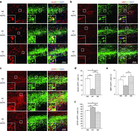
Figure 2.
Differentiation of engrafted neural stem/progenitor cells (NS/PCs) cotransplanted with M0, M1, and M2 macrophages into the intact spinal cord. (a–c) Representative images of NS/PCs derived from GFP-Tg mice and cotransplanted with M0, M1, or M2 macrophages. The NS/PCs were differentiated into (a) NeuN-, (b) MBP-, and (c) glial fibrillary acidic protein (GFAP)-positive cells and detected by IHC. The boxed area in each image is enlarged at the lower left-hand corner of the panel. Space bars = 50 μm. (d–f) The percentages of (d) NeuN-, (e) MBP-, and (f) GFAP-positive cells among the engrafted GFP-expressing NS/PCs are shown. Data in (a–c) are representative of three independent experiments; data in (d–f) were pooled from three independent experiments (*P < 0.05, **P < 0.01).

