Figure 7.
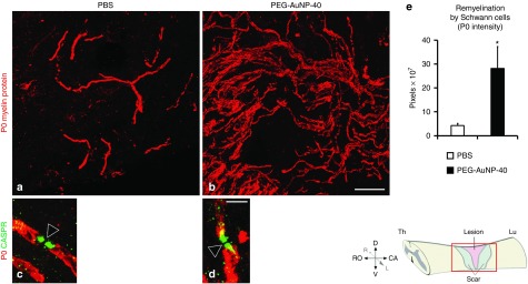
Spontaneous remyelination by Schwann cells is enhanced in mice treated with PEG-AuNP-40, 8 weeks after spinal cord injuries. (a,b) Immunofluorescence labeling and confocal analysis of parasagittal sections of the lesioned spinal cord from mice of the phosphate-buffered saline (PBS) control group (a) or the PEG-AuNP-40 group (b) stained for the Schwann cell myelin marker P0 (red). A notable increase in P0+ myelin internodes is apparent in the PEG-AuNP-40 group compared to the PBS group. (c,d) At high power magnification, double immunofluorescence labeling for P0 (red) and the paranodal marker CASPR/paranodin (green) shows that newly formed myelin paranodal areas (green) are reformed after injury and flank the nodes of Ranvier (empty arrowheads). Scale bar, 40 μm for a, b in b; 5 μm for c, d in d. (e) Quantification of total Schwann cell myelin was performed by measuring P0 immunofluorescence intensity showing an increase in the PEG-AuNP-40 group (n = 6) as compared to the PBS control group (n = 6). *P ≤ 0.05, by Student's t-test. Values represent means ± SEM.

