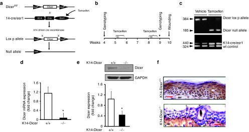
Figure 2.
Conditional deletion of Dicer in mouse epidermis. (a) Deletion of Dicer gene flanked by LoxP sites (Dicerfl/fl) was induced using a tamoxifen-inducible Cre recombinase protein fused to estrogen-receptor (ER) ligand-binding domain under keratin 14 promoter (K14-CreER). When crossed with strain containing Dicerfl/fl, the offspring produces tamoxifen-induced, Cre-targeted keratinocyte-specific deletion of Dicer. (b) Experimental setup and tamoxifen treatment protocol. K14-CreER Dicerfl/fl mice were injected (i.p) once daily for 5 consecutive days with either tamoxifen (80 mg/kg) or vehicle (corn oil) on weeks 5 and 8. On week 9, genotyping was performed followed by wounding on week 10. (c) PCR showing presence of Dicer-null fragments in tamoxifen-treated animals. (d) Epidermis and dermis were separated using dispase digestion. Dicer mRNA expression in epidermis of vehicle- (Dicer+/+) or tamoxifen-treated (Dicer-/-) mice. (e) Representative western blot of Dicer protein in vehicle- or tamoxifen-treated epidermal tissue. The quantification of the signal was normalized by GAPDH, and the result was expressed as mean ± SD (n = 4; *P < 0.001). (f) Immunohistochemical localization of Dicer in the epidermis. Counterstaining was performed using hematoxylin. The dermal–epidermal junction is indicated by a dashed black line in each panel. Bar = 20 μm.

