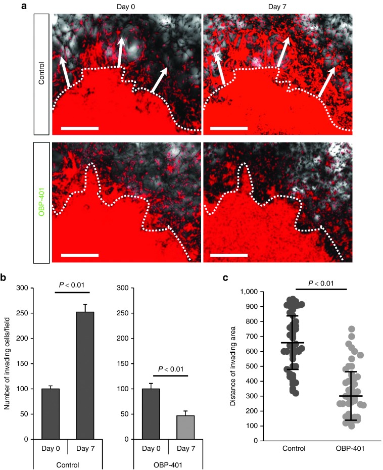
Figure 5.
OBP-401 inhibits invading glioma cells and kills them in 3D Gelfoam histoculture. U87MG cells (5 × 10⁶) expressing RFP, were seeded on Gelfoam for 3D histoculture. OBP-401 was added at 2 × 10⁸ PFU 48 hours after seeding. All images were acquired with the FV1000 confocal laser scanning microscope. (a) Representative images from the RFP channel of mock-infected invading glioma cells (upper) and OBP-401-infected invading glioma cells (lower) cultured in Gelfoam histoculture. Dotted line separates invading area and tumor. Arrows show the direction of invading glioma cells. (b) Histogram shows the number of invading glioma cells for mock-infected and high-dose OBP-401-infected glioma cells in Gelfoam histoculture. (C) Scattergram shows the distance (μm) of invading glioma cells in Gelfoam histoculture from mock-infected glioma cells and high-dose OBP-401-infected glioma cells. Data are shown as average ± SD. n = 5.

