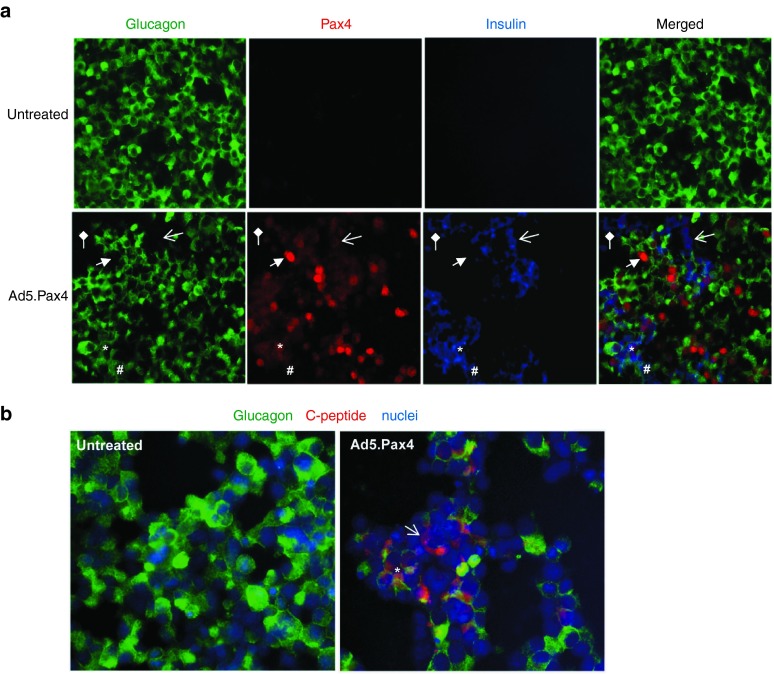
Figure 2.
Pax4-induced insulin and C-peptide expression in αTC1.9 cells. (a) Expression of glucagon (green), Pax4 (red), and insulin (blue) in untreated or Ad5.Pax4 treated cells. The insulin antibody was generated from guinea pig (Abcam, Cambridge, MA), and Pax4 and glucagon antibodies were the same as described in Figure 1. Shown are representative images from 5 days postinfection. The star (*) marks an example of glucagon+/ Pax4+/ Insulin+ (Pax4-positive bi-hormonal) cells. The open arrow marks an example of glucagon−/ Pax4+/ Insulin+ cells, in which glucagon was completely turned off and insulin expression was turned on in these cells. The block arrow marks an example of glucagon− /Pax4+ /Insulin− cells, in which glucagon expression was turned off, but insulin was not induced. The # marks a bi-hormonal cell with barely detectable Pax4, and the diamond arrow marks an insulin+ cell without detectable Pax4. (b) Expression of glucagon (green) and C-peptide (red). The rabbit anti-C-peptide antibody from Cell Signaling Technology (Danvers, MA) was used. Nuclei staining (blue) was used to mark all cells. The open arrow marks an example of glucagon−/ C-peptide+ cells (glucagon is totally inhibited), and the star (*) marks an example of glucagon+/ C-peptide+ cells (bi-hormonal cells).

