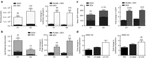
Figure 4.
Glucocorticoid (GC)-priming effect with multiple reporter genes and delivery vehicles, and in the presence of a GC receptor antagonist. The graphs on the left for (a–c) represent transgene expression magnitude for three different reporter genes (a) Luciferase, (b) B-Galactosidase (B-gal), and (c) green fluorescent protein (GFP) intensity as quantified by flow cytometry. Error bars for luciferase expression (a) and B-gal (b) represent standard error (SE) of the mean for triplicate wells collected on duplicate days, while error bars for GFP expression (c) represent SE of the mean for pooled triplicate wells collected on triplicate days. All reporter plasmids show statistically increased expression in GC-primed cells compared to the EtOH control. The graphs on the right for (a–c) show the complete knockdown of the GC-priming effect on both (a and b) transgene expression and (c) transfection efficiency by the addition of known GC receptor antagonist Mifipristone (RU486), represented as fold change of GC primed cells vs. cells treated with both GC and RU486. (d) GC-priming enhances transgene expression using three of the most common commercially available nonviral vehicles, 25 kD branched polyethylenimine, Lipofectamine-2000, and Lipofectamine-LTX, with significantly increased expression of 3.5- to 5-fold, 5- to 6.75-fold, or 8- to 12-fold, respectively, when compared to cells not primed with GCs.

