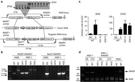
Figure 1.
Generation of hESCs-WASKO cell lines by targeting the WAS locus with ZFNs. (a) Scheme depicting the strategy followed to mutate the WAS gene. Upper diagram illustrates the first five exons of the WAS locus (E1–E5), the sequences recognized by the ZFN pair (left ZFN and right ZFN, in the gray box), the homologous sequences used to promote homologous recombination (H.arm5' and H.arm3') and the primer pair used to identify the wild-type locus (rendering a 2-kb fragment). The middle diagram illustrates the donor DNA used to disrupt WAS expression. Exon 1 has a deletion in the ATG codon to block translation (E1*). Exon 2 includes mutations in the splicing acceptor site (E2*). A neo expression cassette (SV40-neo-pA) has been introduced to allow antibiotic selection. Lower diagram shows the ‘edited' WAS locus after homologous recombination with the donor DNA. Arrows indicate the primers used to identify those clones modified by homologous-directed recombination (HDR). (b) Successful gene editing of AND-1_WT cells. K562 cells wild-type (WT) and K562-WASKO (KO)41 have been used as negative and positive control of gene editing, respectively. Agarose gel shows the analysis to detect HDR in two G418-ressistant clones of the hESC line AND-1_WT. The band of 3.5 kb, present in both G418-resistant clones as well as in the K562 WASKO cells, indicates successful HDR. Also, the presence of the 1.9 Kb band in AND-1_WASKO as in the positive control indicates that the HDR happened in the right orientation. (c) WAS expression was abolished upon hematopoietic differentiation in AND-1_WASKO cells. WAS and CD45 gene expression analysis performed by RT-qPCR of AND-1_WT (WT) and AND-1_WASKO clones (KO1 and KO2) before (ND) and after (D) 15 days of hematopoietic differentiation. CD45 expression indicates the hematopoietic specification. As expected WAS expression was not detected in AND-1_WASKO clones. Values are mean of three experiments ± standard error of the mean. (d) Absence of WASp protein in hematopoietic differentiated AND-1_WASKO cells. Western blot analysis of protein extracts from AND-1_WT and AND-1_WASKO cells, obtained at days 8 (D8) and 15 (D15) of the hematopoietic differentiation. The membrane was hybridized with D1 (specific for WASp) and ERK (as loading control) antibodies. WASp protein was not detected in any of the AND-1_WASKO clones. K562 cells wild-type (WT) and K562-WASKO (KO) have been used as negative and positive control for WASp expression.

