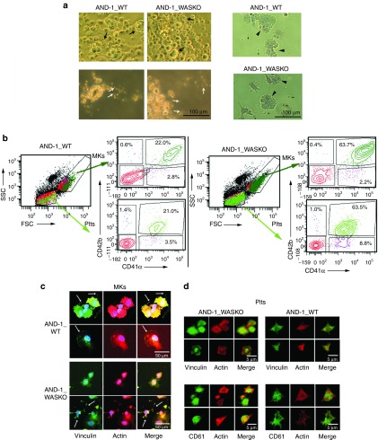
Figure 4.
Megakaryocytic differentiation and Plts production of hESCs-WASKO cells are not impaired. (a) Morphological analysis of megakaryocytic differentiation cultures of AND-1_WT1 and AND-1_WASKO_c1.1 cells (see Supplementary Figure S5 and Materials and Methods for details). Day 20 megakaryocytic cultures were photographed with a microscope Primo Vert (Carl Zeiss) on tissue culture plates (top-left panels), after collection (bottom-left panels) or after adhesion to fibrinogen coated slides and stained with Papanicolau (right panels). MKs and Plts-like structures are indicated by arrows. (b) Phenotypic characterization of cells contained in the megakaryocytic cultures. Cells were gated based on size (FSC) and granularity (SSC) into MK-region and Plts region (SSC/FSC plots). Each gate was further analyzed for expression of CD41 and CD42 (right plots at the side of the FSC/SSC plot). Large and granular, CD41+CD42+ cells were identified as MKs (SCChighFSChighCD41+CD42+; top plots, dark green). Small CD41+CD42+ cell fragments were identified as Plts (SSClowFSClowCD41+CD42+; bright green). (c) MK derived from AND-1_WT1 and AND-1_WASKO bind to fibrinogen and express CD61 and vinculin. Megakaryocytic cultures from AND-1_WT1 (top panels) and AND-1_WASKO_c1.1 (bottom panels) were collected on day 20, adhered to fibrinogen coated slides, activated with thrombin and immune-stained Phalloidin (red, actin), anti-vinculin (green) and 4',6-diamidino-2-phenylindole (DAPI) (blue). A Merge image is shown at the right of each set. (d) Platelets from AND-1_WT1 (top panels) and AND-1_WASKO_c1.1 (bottom panels) were collected on day 20 and treated as above. Samples were immune-stained with DAPI (Blue), Phalloidin (red) together with anti-vinculin (green) (top panels) or anti-CD61 (green) (bottom panels). A Merge image is shown at the right of each set. Confocal images were captured with a confocal microscope Zeiss LSM 880.

