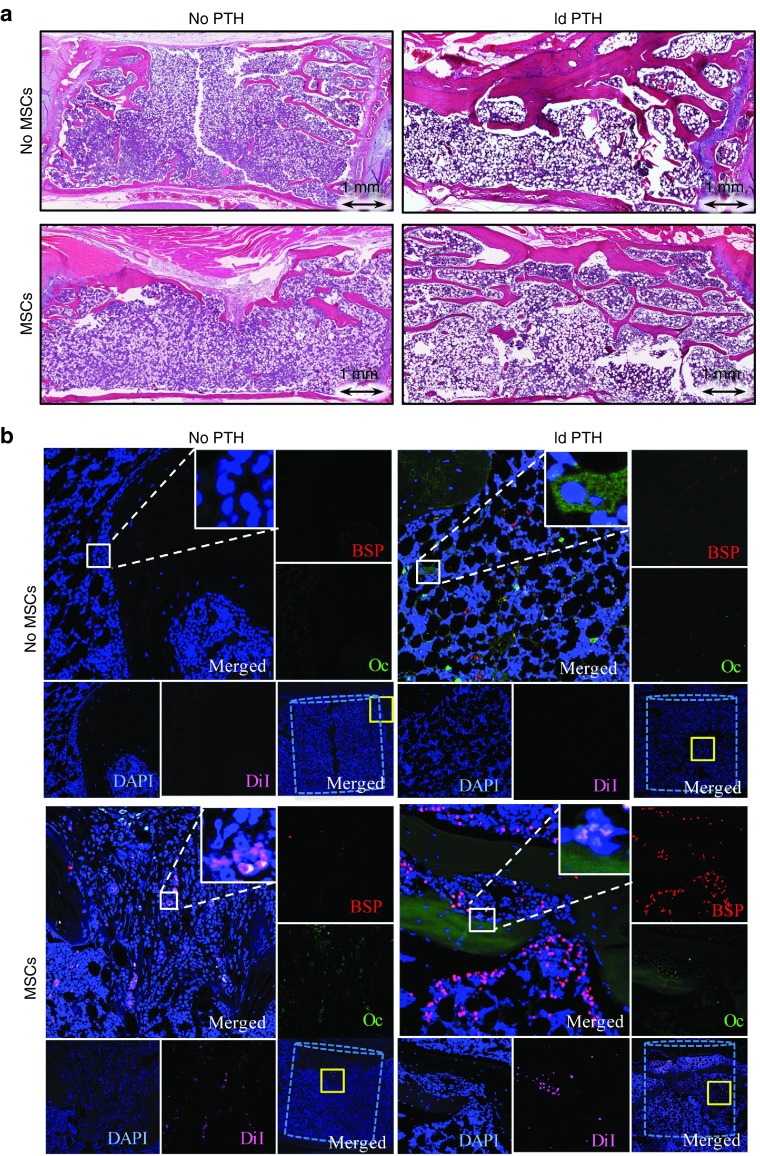
Figure 4.
Intravenous mesenchymal stem cells and parathyroid hormone therapy regenerates vertebral defects in osteoporotic rats: histological and immunofluorescence analyses. (a) Representative vertebral defects of animals that received low-dosage PTH (ldPTH) or vehicle and DiI-labeled mesenchymal stem cells (MSCs) or saline were harvested, decalcified, embedded in paraffin and sectioned. Slides were stained with standard hematoxylin and eosin and imaged with light microscopy. (b) Serial slides were also immunostained against the osteogenic markers osteocalcin (Oc), bone sialoprotein (BSP), and counterstained with DAPI. Slides were imaged using confocal microscopy for DAPI, DiI-labeled MSCs, Oc, and BSP. A representative panel for each treatment group includes: (i) a merged image of the defect site with the bone void marked by a dashed cylinder and an area subsequently magnified marked by a solid yellow square (bottom right corner); (ii) a merged magnification of the area denoted by the yellow square (top left corner) with a built in magnification of the solid white square in its' top right corner; (iii) four single channel magnifications of the solid yellow square-marked area along the right and bottom edges denoted: BSP, Oc, DAPI, and DiI corresponding to the stain in the subfigure. DAPI, 4',6-diamidino-2-phenylindole dihydrochloride.

