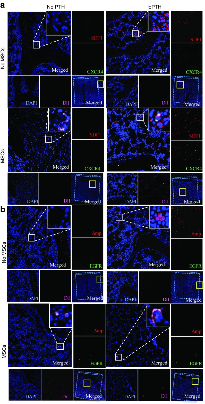
Figure 5.
Parathyroid hormone enhances migration of mesenchymal stem cells to the defect site via two pathways: confocal imaging of immunofluorescent staining. Representative vertebral defects of animals that received low-dosage PTH (ldPTH) or vehicle and DiI-labeled mesenchymal stem cells (MSCs) or saline were harvested, decalcified, embedded in paraffin, and sectioned. Slides were immunostained against migration markers (a) stromal cell-derived factor 1 (SDF1) and C-X-C chemokine type 4 (CXCR4) or (b) epidermal growth factor receptor (EGFR) and amphiregulin (Amp) and both sets counterstained with DAPI. Slides were imaged using confocal microscopy and a representative panel for each treatment group is shown including: (i) a merged image of the defect site with the bone void marked by a dashed cylinder and an area subsequently magnified marked by a solid yellow square (bottom right corner); (ii) a merged magnification of the area denoted by the yellow square (top left corner) with a built in magnification of the solid white square in its' top right corner; (iii) four single channel magnifications of the solid yellow square-marked area along the right and bottom edges named corresponding to the stain in the subfigure. DAPI, 4',6-diamidino-2-phenylindole dihydrochloride.

