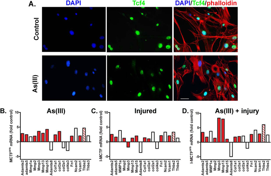
Figure 4.
Fibroblast extracellular matrix (ECM) transcript expression in recovery. MCT fibroblasts were isolated from control, injured, As(III)-exposed, or As(III)-exposed and injured mice. More than 90% of the cells were positive for Tcf4, a transcription factor of fibroblasts (A; Scale bar = 50 µm). (B–D): Cells were cultured for two passages in the absence of As(III) before RNA was isolated and transcripts were measured with an ECM-targeted quantitative polymerase chain reaction array. Transcript abundance was normalized to a suite of house-keeping genes and the graphs present the fold increase in transcripts in cells isolated from As(III)-exposed mice (B), injured mice (C), and As(III)-exposed, injured mice (D), relative to transcript abundance in uninjured, control mice. Red bars are NF-κB driven genes and the striped bar codes for Thbs2 that stimulates NF-κB. Abbreviations: DAPI, 4’,6-diamidino-2-phenylindole.

