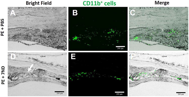
Figure 6.

Immunohistochemical staining macrophages with mouse FITC-labeled anti-CD11b antibodies. CD11b+ cells were scattered in the marrow cavity caused in PBS group; however, for 7ND-treated group, all CD11b+ cells localized in bone marrow cavity. Original magnification = ×200, green: FITC-labeled anti-CD11b, white arrow: bone marrow cavity.
