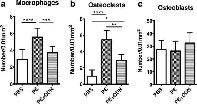
FIGURE 3.
Cell numbers in the calvarial immunostaining sections. (a and b) macrophages and osteoclasts were detected using murine anti CD11b and TRAP staining, respectively. The number of these cells was reduced with the administration of NFκB decoy ODN in the presence of PE particles. (c) The number of osteoblasts immunohistochemically stained using anti-mouse ALP showed no significant differences among the groups. *, p < 0.05; **, p < 0.01; ***, p < 0.001; and ****, p < 0.0001.

