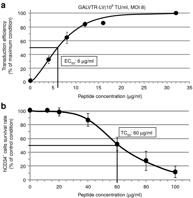
Figure 2.
Determination of the half maximal efficient (EC50) and toxic (TC50) concentrations of Vectofusin-1. (a) hCD34+ cells were infected with GALVTR-LVs in the absence or presence of various concentrations of Vectofusin-1. Transduction efficiencies (percentage of GFP+ cells) were obtained 5 days post-transduction (n = 4). Data are normalized to the maximum effect observed ± SD (average maximal value of transduction was 67%). (b) Evaluation of the TC50 of Vectofusin-1. The hCD34+ cells were incubated overnight with the indicated amounts of Vectofusin-1 (n = 6). The survival rate was estimated by counting the number of living cells using the Trypan blue exclusion method under light microscopy. Data are normalized to the control condition ± SD (average value of survival rate in the absence of peptides was 98.9%). GALV, gibbon ape leukemia virus; LV, lentiviral vector; MOI, multiplicity of infection; TU, transducing unit.

