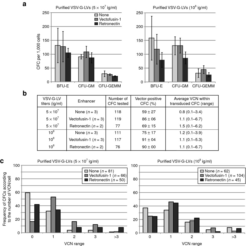
Figure 4.
Lack of in vitro hematopoietic toxicity of Vectofusin-1. (a) Differentiation of transduced hCD34+ cells in colony-forming cell (CFC) assays. Results represent the average number of different types of colonies obtained for 1,000 cells plated after transduction with 5 × 10⁷ and 108 ig/ml (conditions of Figure 3). (b) Transduction was measured in individual CFCs obtained after 2 weeks of culture in methylcellulose. Transduction of CFCs was measured by determining the percentage of GFP+ CFCs using epifluorescence microscopy (“vector-positive CFC (%)”), and the vector copy number (VCN) in each CFC by quantitative PCR. The average VCN per vector-positive CFC and range are listed (right column). (c) Bars represent the percentage of CFCs in each category (VCN range) over the total number of CFCs analyzed. The number of CFCs analyzed is indicated between brackets for each group. BFU-E, burst-forming unit, erythroid; CFU-GEMM, colony-forming unit, granulocyte, erythrocyte, macrophage, megakaryocyte; CFU-GM, colony-forming unit, granulocyte-monocyte; GFP, green fluorescent protein; ig, infectious genome; VSV-G-LV, vesicular stomatitis virus-G-lentiviral vector.

