
Fig 1. Corneal whole mount imaging.
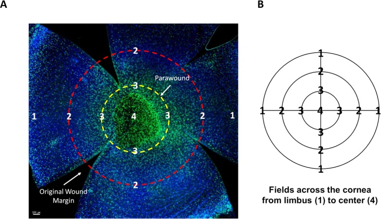
(A) Example of a whole mount, 18 hours post-wound, stained with Ly6G (green) to visualize infiltrating neutrophils and DAPI (blue). Radial cuts divide the cornea into four petals, enabling the cornea to flatten under the coverslip. The wound area is visible in the central cornea surrounded by the parawound region (yellow). The location of the original wound margin is demarcated (red). Scale bar = 100μm (B) Each cornea was measured from the limbus across the center to the opposite limbal region using x- and y- image coordinates and images were taken at even intervals, designated by points 1 through 4.