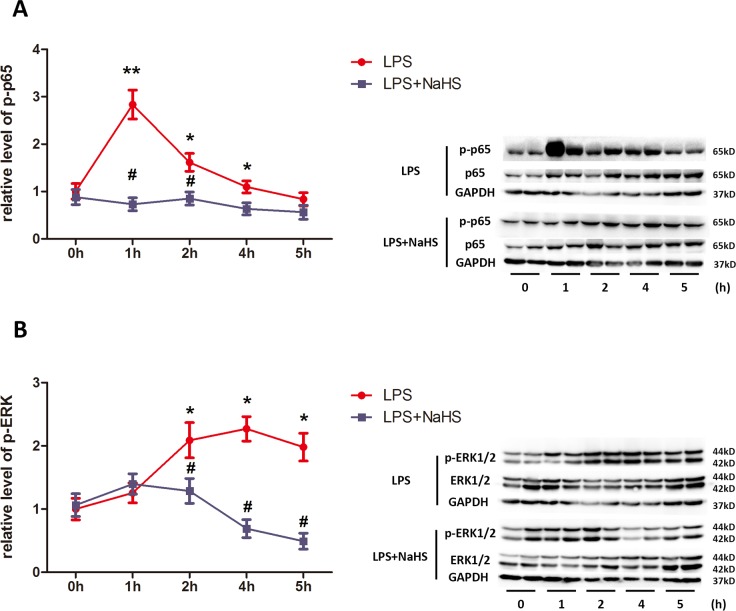
Fig 6. NaHS attenuates LPS-induced activation of ERK1/2 and p65 in myometrium.
The uterine tissues were harvested 1h, 2h, 4h and 5h after LPS and/or NaHS injection. The levels of phosphorylated p65, p65, phosphorylated ERK1/2 and ERK1/2 were determined by western blotting. Phosphorylated p65 (A) and ERK1/2 (B) were normalized by p65 and ERK1/2. Values are presented as mean ± SEM. N = 4. * P<0.05, **P<0.01 compared with vehicle control group, #P<0.05, ##P<0.01 compared with LPS-injected group.

