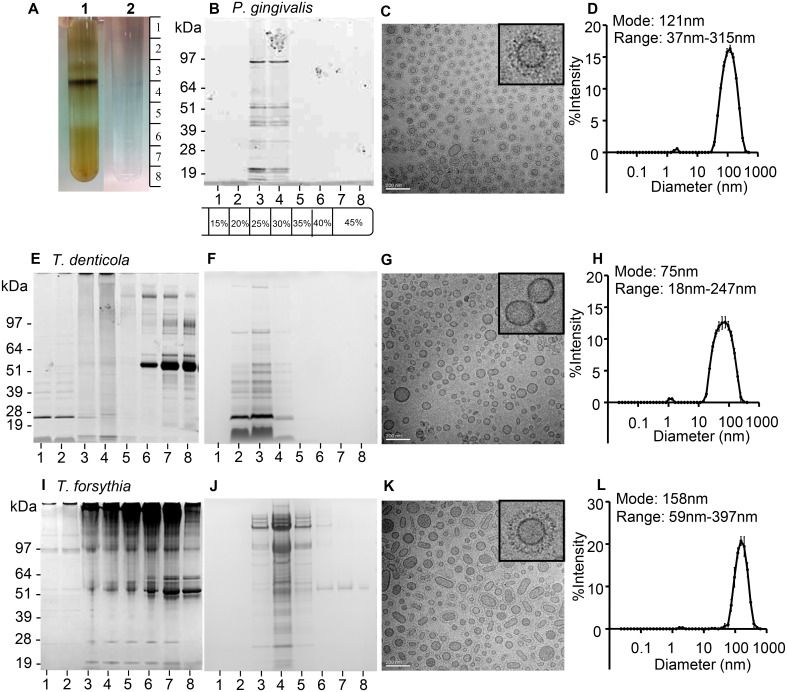
Fig 1. Isolation of highly purified P. gingivalis, T. denticola and T. forsythia OMVs.
OptiPrep density gradient centrifugation of a crude T. forsythia OMV preparation (A1) separated non-OMV associated material (bottom of density gradient) from highly purified OMVs isolated at a higher density. A blank OptiPrep density gradient is shown in A2. SDS-PAGE of P. gingivalis (B), T. denticola (E F) and T. forsythia (I J) Optiprep density gradient fractions taken in eight 1.5 mL gradient fractions from top to bottom following centrifugation. Gradient fractions were subjected to SDS-PAGE and protein bands visualized by SimplyBlue or SyproRuby staining. Molecular mass markers (Novex SeeBlue Plus2 Prestained Standard) are indicated in kDa. OMVs were observed using transmission electron microscopy (TEM) (C G K). Vesicle size and range was further confirmed using dynamic light scattering (DLS) on pooled and washed OMV containing gradient fractions (D H L).

