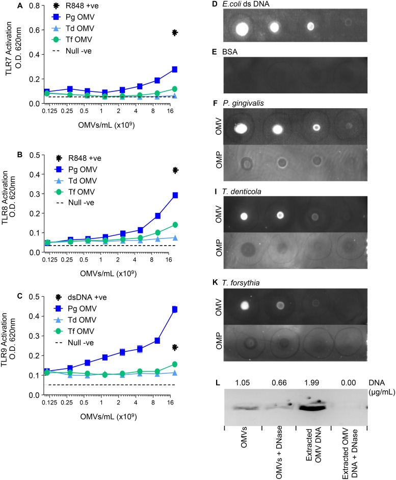
Fig 4. TLR7, TLR8 and TLR9 Activation by P. gingivalis, T. denticola and T. forsythia OMVs.
HEK-Blue TLR7 (A), TLR8 (B) and TLR9 (C) Cell lines were challenged with 20μL of either purified OMVs or ligands R848 (10 μg/mL in 2 fold dilutions) and dsDNA (100 μg/mL in 2 fold dilutions) respectively. Alkaline phosphatase secretion was determined after 20 hours incubation at 620nm on a spectrophotometer. Monoclonal antibody αdsDNA MAB030 was used to detect DNA in purified OMV preparations and Triton X-114 extracted Outer Membrane Protein (OMP) preparations, used at 0.5 mg/mL protein and 1.5 mg/mL protein respectively, for P. gingivalis (F), T. denticola (I) and T. forsythia (K). OMV preparations were spotted on a nitrocellulose Immuno-Blot PVDF Membrane for Protein Blotting in three 10-fold dilutions. E. coli double stranded DNA was used as a positive control at 50 ng/mL in three 10-fold dilutions (D). Bovine Serum Albumin was used as a negative control at 0.5 mg/mL in three 10-fold dilutions (E). P. gingivalis whole OMVs and extracted OMV DNA were treated with DNase and DNA concentrations determined using Qubit Assay Kits and SYBR Safe DNA gel stain (L).

