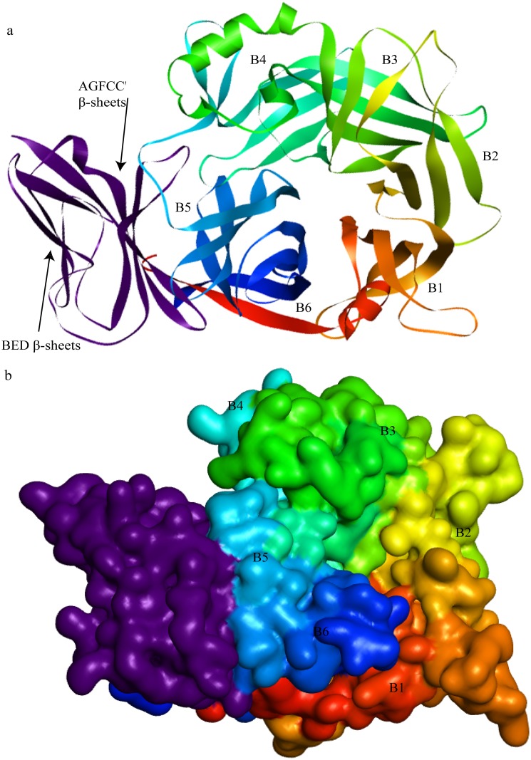
Fig 3. Cartoon and Solvent drawing of predicted PPRVHv-shSLAM complex.
(A)The PPRVH head domain exhibits the six-bladed β-propeller fold (rainbow colors) and forms a monomer. The sheep SLAM (purple) exhibits a typical β-sandwich structure with BED and AGFCC′β-sheets. The groove in the B4 blade and B5 of the head domain of PPRVH bind to the AGFCC′ β-sheets of the membrane-distal ectodomain of shSLAM. (B) The pattern of the PPRV with SLAM.

