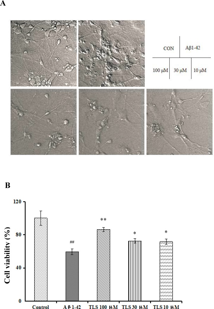
Fig 8. Effects of TLS on cell viability of the primary cultured mouse neuronal cells induced by Aβ1–42.
Primary mouse neurons were treated with TLS (10–100 μM) and Aβ1–42 (10 μM) for 48 h. (A) Morphological characteristics of primary cultured mouse neuronal cells. (B) Cell viability was assessed by MTT assay. #p < 0.05, ##p < 0.01 compared with the control group; *p < 0.05, **p < 0.01 compared with the Aβ1–42 group.

