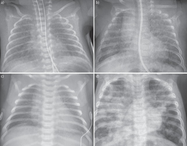
Figure 1.
Chest radiograph images. a) Intubated 23+6 weeks preterm infant with RDS. Note bilateral ground glass shadowing and air bronchograms. The ET tube is low in this image and needs withdrawing. Parental consent obtained for publication. b) Ex 24-week preterm infant with CLD. Note areas of cystic changes and linear shadowing throughout both lungs. Parental consent obtained for publication. c) Term infant with TTN. Note wet silhouette around the heart and fluid in the horizontal fissure. Image: © Auckland District Health Board. d) Term infant with MAS. Widespread asymmetrical patchy shadowing throughout both lungs with hyperinflation. Reproduced from [21] with permission from the publisher.

