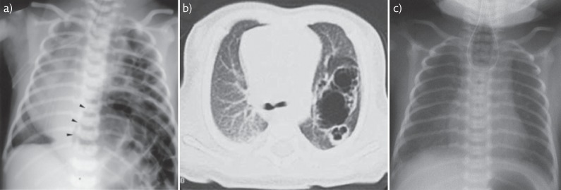
Figure 2.
Radiology images of surgical conditions/congenital anomalies. a) Chest radiograph of infant with large left sided CDH. Note presence of bowel and stomach (arrowheads) within the chest. Mediastinal shift to the right. Reproduced from [91] with permission from the publisher. b) CT image of left sided CCAM demonstrating large cystic areas and c) chest radiograph demonstrating coiled nasogastric tube in the upper oesophageal pouch indicating oesophageal atresia. Note gas in stomach indicating presence of fistula between distal oesophagus and trachea. b) and c) reproduced from [21] with permission from the publisher.

