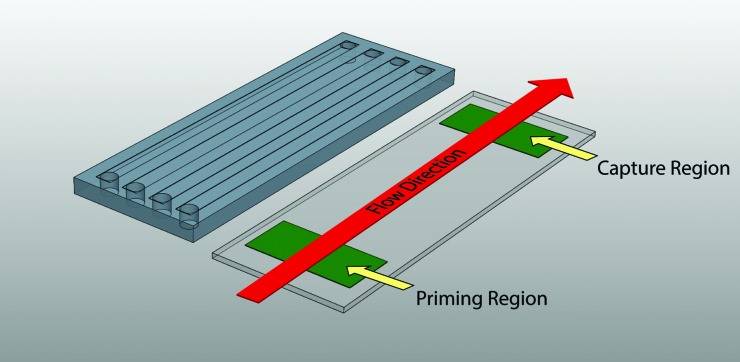
Fig. 1.
Schematic of a flow assay assembly. Protein agonists are deposited on a glass substrate in the priming and capture regions by microcontact printing and covalent attachment of proteins is achieved through the use of a commercial NHS ester chemistry (Nexterion-H®, Schott). A relief-molded PDMS flow channel is inverted on the stamped surface to create the final assembled device. Flow through the device brings blood past the priming region followed by the capture region before exiting the device. Platelets near the substrate surface are therefore able to sequentially interact with two surface-bound agonist regions in a controlled flow environment.

