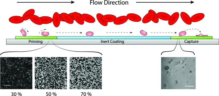
Fig. 2.
Schematic of flow assay function. The presence of red blood cells in flow creates a margination effect which drives platelets toward the chamber walls. An increased number of platelets near the substrate surface relative to the bulk flow increases chances of platelet–surface interactions. Platelets near the substrate transiently roll along the surface and become primed through interactions with agonists. A variable density of printed agonists in the priming region provides a range of probabilities that a platelet contacting the surface in the priming region will interact with a printed agonist. Shown here are fluorescent images of microcontact printed fibrinogen at three different surface coverage densities, 30%, 50%, and 70% (scale bar represents 10 μm). Primed platelets continue to flow downstream along a platelet-inert surface. Primed platelets encountering the downstream agonist patch have an increased propensity to form stable adhesions compared to nonprimed populations. The number of adhered platelets in the capture region is used as an indication of overall priming in the platelet population. The inset image is a representative view of platelets adhered to a fibrinogen capture region (scale bar represents 20 μm).

