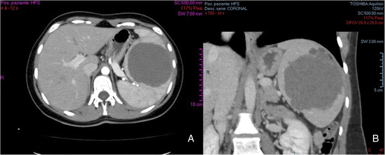
Fig. 1.

Preoperative contrast-enhanced abdominal CT scan: a the axial projection shows the recurrent splenic mass (7.7 × 8.5 cm), disclosing absence of calcifications, no contrast uptake; b the sagittal projection shows the presence of satellite nodules, and the splenic parenchyma almost totally replace
