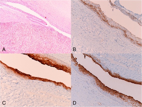
Fig. 2.

Histological examination shows a an epidermoid cyst with fibrous walls and a stratified squamous lining (top) within the spleen parenchyma (bottom) (hematoxylin and eosin, original magnification ×100); b the epithelium is immunoreactive with EMA, c CEA, and d CA19-9 (original magnification ×200)
