
Fig. 2.
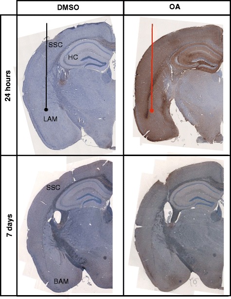
Characterisation of tau pathology. Representative coronal sections from the injection site are stained for phospho-tau with AT180 and counterstained with haematoxylin for cell bodies. Tissue injected with DMSO have relatively low levels of phospho-tau immunoreactivity. At 24 h phospho-tau is abundant at the injection site in the lateral amygdala (LAM) and can be seen throughout the layers of the cortex (CX) and hippocampus (HC) in OA injected. At 7 days phospho-tau persists primarily in the basal amygdala (BAM) and somatosensory cortex (SSC) in the OA injected mice