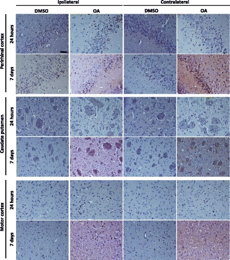
Fig. 4.

Tau phosphorylation propagates to areas anterior to the injection site. Representative images of AT180 immunoreactivty are taken from brain regions 2.08 mm anterior to the injection. At 24 h there is relatively little tau phosphorylation detectable in anterior regions, although a few positive cell bodies in the perirhinal cortex are noted. At 7 days post OA injection tau phosphorylation is pronounced in the perirhinal cortex in both the injected and contralateral hemisphere. In the striatum phospho-tau immunoreactivity is seen confined to within axon bundles. Only a small proportion of phospho-tau positive neurones are observed in the motor cortex. Scale bar = 50 μM
