Fig. 5.

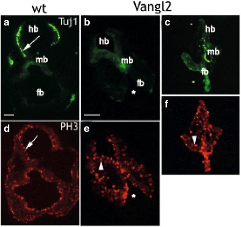
Vangl2 overexpression impairs differentiation during embryonic development. a-f Micrographs of 10 μm transversal tissue sections of neural mouse embryo E9.5 tissue labeled with antibodies against beta-tubulin-III (Tuj1, green) and phospho-Histone-3 (P-H3, red) and visualized with fluorescent conjugated secondary antibodies respectively. Wild-type E9.5 mouse embryos showed an even distribution of Tuj1+ neurons in the hindbrain part of the neuroepithelium (arrow in a), but were almost completely absent in the neuroepithelium of nestin-Vangl2 embryos (b) and (c). The labeling against M-phase marker PH3 was similar in the neuroepithelium of both wild type and transgenic sections (arrow in (d) and arrowhead in (e) and (f). Transgenic E9.5 Vangl2 embryos displayed impaired cranial neurulation (indicated with *[17];). Scale bars are 200 μm
