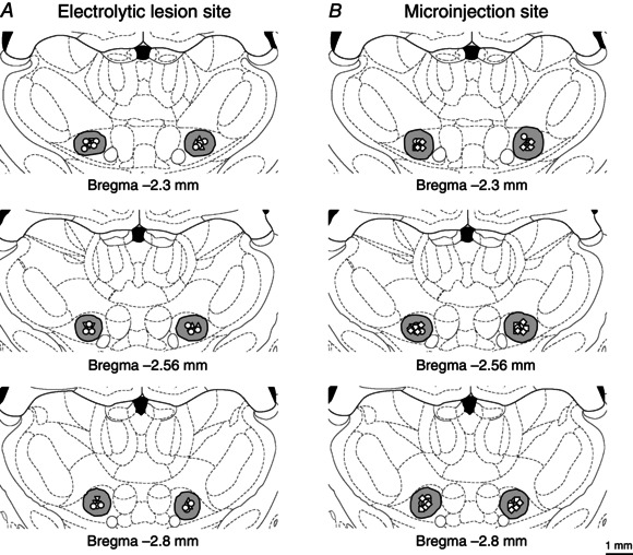
Figure 4. Locations of electrolytic lesions of thalamic ventromedial nuclei and of microinjection sites within the bilateral ventromedial nuclei .

Reconstructions of the locations of 30 electrolytic lesions of thalamic ventromedial (VM) nuclei (A), and of 50 microinjection sites within the bilateral VM nuclei (B). The spread regions of lesion and microinjection are marked by grey areas. Effective sites for ipsilateral, contralateral and bilateral electrolytic lesions are indicated by up‐triangles, down‐triangles and circles in A, respectively. Microinjection sites of 0.9% saline, atipamezole and WAY‐100635 are indicated by circles, squares and diamonds in B, respectively. Note that some microinjection sites overlap with others in this reconstruction.
