
Figure 5. Exercise‐induced specific neuroprotection of fast vs. slow motor neuron subpopulations in type 3 SMA‐like mice is SMN independent .
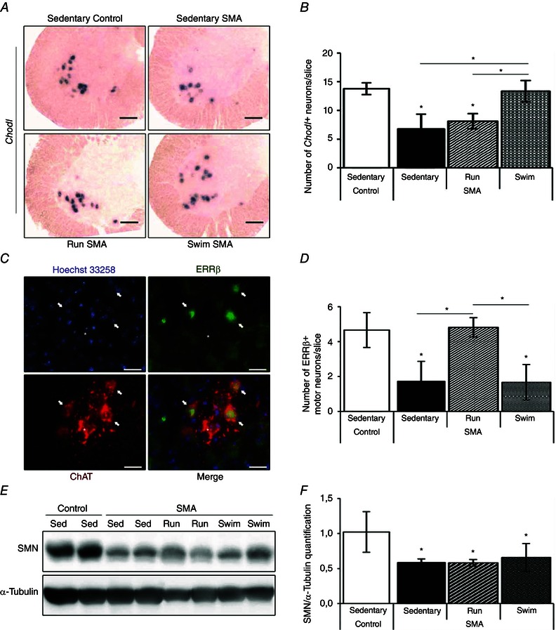
A, in situ hybridization on Chondrolectin mRNA (Chodl) in lumbar spinal cords of sedentary control (top left) and type 3 SMA‐like mice (top right) compared to running‐trained (bottom left) and swimming‐trained (bottom right) type 3 SMA‐like mice at 12 months of age (scale bar: 100 μm). B, quantitative analysis of the number of Chodl‐positive neurons in the ventral horn of lumbar spinal cords of sedentary control compared to sedentary, running‐trained (Run) and swimming‐trained (Swim) type 3 SMA‐like mice at 12 months of age (n = 4 in each group). C, immunodetection of the slow‐type motor neuron marker oestrogen‐related receptor β (ERRβ, green) in the nucleus (Hoechst 33258, blue) of ChAT‐positive motor neurons (ChAT, red) in the lumbar spinal cords of sedentary type 3 SMA‐like mice at 12 months of age (scale bar: 25 μm). D, quantitative analysis of the number of ERRβ‐positive motor neurons in the ventral horn of lumbar spinal cords of sedentary control mice compared to sedentary, running‐trained (Run) or swimming‐trained (Swim) type 3 SMA‐like mice at 12 months of age (n = 4 in each group). E and F, Western blot analysis (E) and quantification (F) of SMN protein expression in the ventral lumbar spinal cord of sedentary control and type 3 SMA‐like mice (Sed) compared to running‐trained (Run) or swimming‐trained (Swim) type 3 SMA‐like mice at 12 months of age (n = 4 in each group). Data are represented as means ± SD (*P < 0.05).