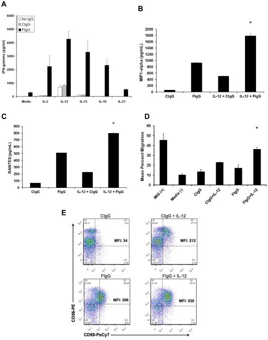
Figure 4. Human NK cells activated in response to F‐IgG‐coated KB cells and cytokines secrete IFNγ, MIP-1α, and RANTES and become motile.
(A) NK cells were cocultured with F-IgG–coated KB tumor (FR-positive) cells in the presence of IL2, IL12, IL15, IL18, or IL21. Control conditions consisted of media only or C-IgG–coated tumor cells in the presence of the cytokines. Culture supernatants were harvested after 48 hours and analyzed for (A) IFNγ, (B) MIP‐1α, and (C) RANTES by ELISA. Ccombinatorial treatment resulted in significant cytokine/chemokine production (P < 0.0001). (D) T cell chemotaxis was assessed with coculture supernatants. MIG treatment of media served as a positive control for T cell migration. (*) Indicates significant enhancement as compared to all other treatment groups except MIG (P < 0.01). (E) Activated NK cells were assessed via flow cytometric analysis for CD56 and CD69. Results are representative of data derived from n ≥ 2 donors.

