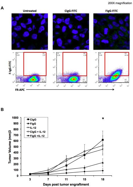
Figure 6. F-IgG binds to KB cell in vivo and IL12 enhances its antitumor effects in a murine model.
(A) Mice bearing KB tumors were treated with 100 mg/kg CIgG‐FITC or FIgG-FITC and sacrificed after 72 hours. Tumors were harvested and fixed/frozen or dissociated. FITC content (green) was assessed in fixed/frozen tissues via confocal microscopy (top panels). Nuclei are stained blue with DAPI (200X images). FR status and FITC content were then measured via flow cytometric analysis (bottom panels). Dual positive FR+/FITC+ cells indicate specific binding of F-IgG to FR+ tumor cells. (B) Mice bearing subcutaneous L1210JF tumors were treated once every 4 days with 100 mg/kg murine C-IgG, 100 mg/kg murine F‐IgG, 2.5 ng murine IL12, C-IgG plus IL12 or F‐IgG plus IL12. Data is representative of n = 3 independent experiments. No significant differences in tumor volume were found between the five groups at baseline. However, at 18 days after treatment, the average tumor volumes of mice receiving either IL12 or F-IgG alone were smaller than those of the C‐IgG‐treated mice (*P ≤ 0.05).

