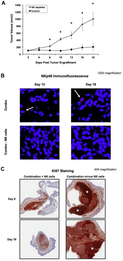
Figure 7. The antitumor effects of F-IgG plus IL12 are dependent on NK cells and result in decreased tumor cell proliferation.
(A) Mice bearing subcutaneous L1210JF tumors were depleted of NK cells with an antibody to the asialo moiety, or treated with an istoype control antibody. n ≥ 4 mice/group were treated with F-IgG (100 mg/kg) and IL12 (2.5 ng). *P ≤ 0.05. (B) Confocal microscopy (120X) detected activated NK cells (NKp46+ red stain) only in nondepleted mice (top, white arrow). Tumor sections were counterstained with DAPI for nuclear identification (blue). (C) Mice were sacrificed at the indicated time points and whole tumors were harvested and sectioned to measure tumor cell proliferation by Ki67 staining (5X). Scale bar indicates 800 um. White asterisks indicate areas of high Ki67 staining within the tumor. Data is representative of n = 3 independent experiments.

