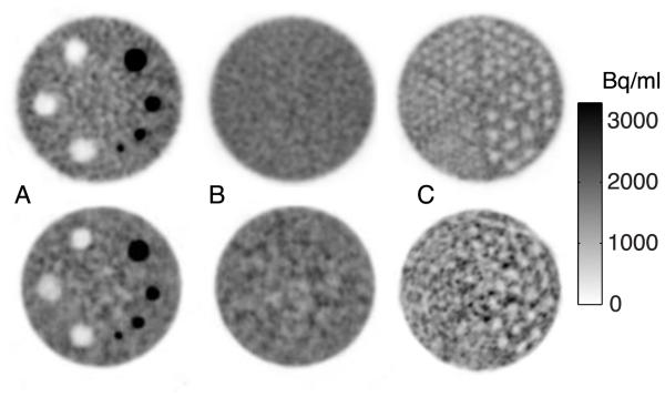
FIGURE 4.
NeuroPET/CT (top) and HR+ (bottom) ACR-type phantom showing 10 mm transverse slices of the (A) contrast section with cold and hot cylinders (B) uniform section and (C) cold rod section for resolution, with the second smallest (6.4 mm) rods resolved for the NeuroPET/CT and the third largest (9.5 mm) rods resolved for the HR+, although lower resolution in the HR+ could be due to the proximity to the edge of the axial FOV. ACR: American College of Radiology

