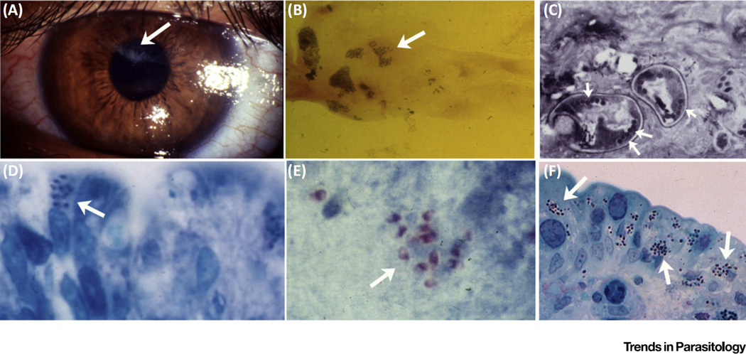
Figure 1. Microsporidiosis in Humans.
(A) Encephalitozoon hellem keratoconjunctivitis. Areas of corneal damage due to microsporidiosis (arrow). (B) Corneal scraping from a case of microsporidian keratoconjunctivitis demonstrating spores (arrow) of E. hellem. (C) Conjunctival biopsy in a case of microsporidian keratoconjunctivitis demonstrating microsporidian spores in cross-section (arrows point to polar tubes, the infective structures). The arrangement of the polar tubes is consistent with Encephalitozoon. (D) Intestinal biopsy from a patient with gastrointestinal microsporidiosis and diarrhea due to Enterocytozoon bieneusi (arrows point to spores in the apical region of an intestinal epithelial cell). (E) Stool stained with modified trichrome stain (arrows point to spores). PCR confirmed that this infection was due to E. bieneusi. (F) Intestinal biopsy from a patient with gastrointestinal microsporidiosis and diarrhea due to Encephalitozoon intestinalis.

