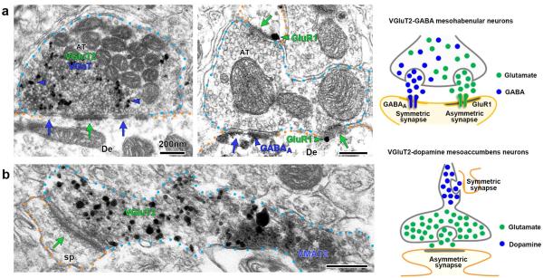
Figure 2. Ultrastructural immunolabeling reveals unpredicted mechanisms of neurotransmission within the mesohabenular and mesoaccumbal pathways.
(a) Lateral habenula micrograph (left panel) showing a single mesohabenular axon terminal containing VGluT2 (scattered dark material detected by immunoperoxidase labeling) and VGaT (gold particles detected by immunogold; blue arrowheads). This single axon terminal forms both an asymmetric synapse (green arrow) and a symmetric synapse (blue arrow) with a common postsynaptic dendrite (De). Postsynaptic to a single axon terminal (middle panel), GluR1 receptors (green arrowhead) are found adjacent to asymmetric synapses (green arrows), while GABAA receptors (blue arrowhead) are found adjacent to symmetric synapses (blue arrow). (b) Nucleus Accumbens micrograph showing a messoaccumbal axon containing both VMAT2 (scattered dark material) and VGluT2 (gold particles). VMAT2 and VGluT2 are segregated within the same axon. Note that the VGluT2 microdomain corresponds to an axon terminal establishing an asymmetric synapse (arrow) with a postsynaptic dendritic spine (sp). All scale bars represent 200 nm.

