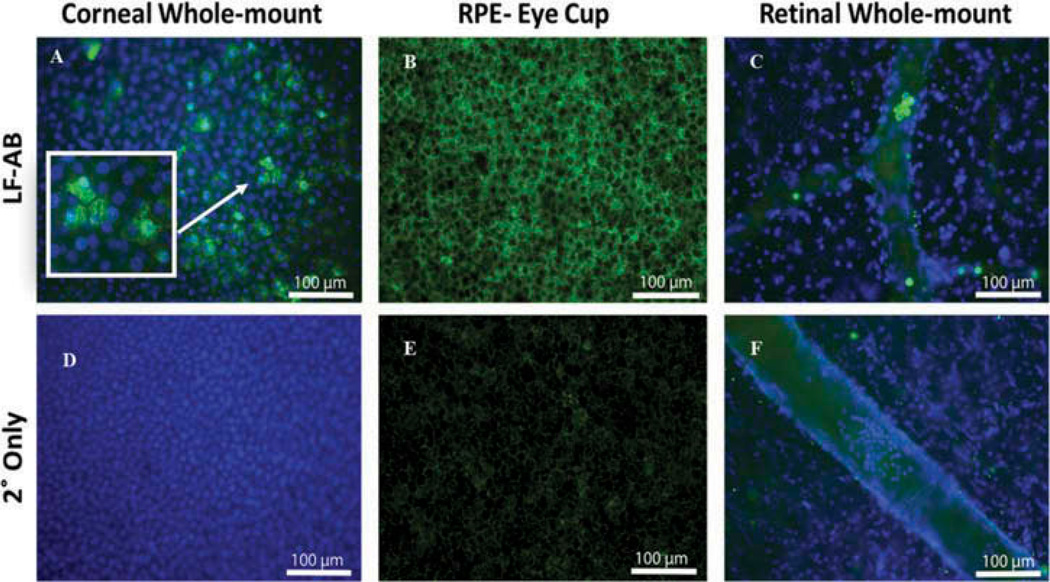
Figure 2.
Immunohistochemistry of flat mounts of cornea, RPE and retina from a human donor eye. (A) LF (ab166803) staining (green) in human cornea with DAPI-stained nuclei (blue). Arrows point to cells with nuclear and cytoplasmic staining. Box shows enlarged image of cells. (B) LF staining in human RPE. (C) LF staining in human retinal tissue counterstained with DAPI to mark nuclei. There is a positive LF staining present only in cells within blood vessels. (D) Secondary only staining of corneal epithelium cells. (E) Secondary only staining of RPE cells. Background fluorescence is caused by the lipofuscin autofluorescence. (F) Secondary only staining of retina containing a retinal blood vessel. *Green, LF; blue, DAPI; arrow, blood cells in C and F.

