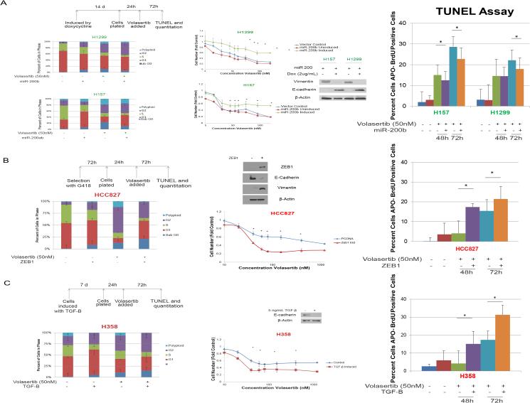
Figure 5.
Mesenchymal cells are more sensitive to PLK1 inhibition than are epithelial cells in isogenic human NSCLC models. A, forced expression of miR-200 led to increased E-cadherin expression and decreased vimentin expression according to Western blotting and to volasertib resistance according to an MTT assay. Induction of a mesenchymal phenotype by ZEB1 expression (B) or 5 ng/mL transforming growth factor-β (C) led to volasertib sensitivity and increased volasertib-induced apoptosis. *P ≤ 0.05 compared with a control or as indicated. PLK1 inhibition-sensitive and -resistant cell lines are indicated in green and red, respectively.

