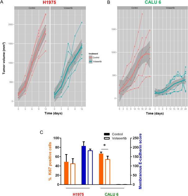
Figure 6.
PLK1 inhibition leads to decreased mesenchymal NSCLC tumor growth in vivo. Mice bearing epithelial (H1975; A) or mesenchymal (Calu6; B) NSCLC tumors were given 30 mg/kg volasertib intravenously each week or a vehicle control. Tumors were measured every 3 days. Individual tumors are graphed as thin lines with markers. The mean tumor size is indicated by the thick solid line, with the standard deviation indicated in dark gray. (C) Tumors were subjected to immunohistochemical staining with Aperio digital analysis. The average percent of tumor cells positive for Ki67 (orange bars) and the average score for E-cadherin (blue bars) were calculated with error bars representing standard deviation. Open bars, volasertib. Solid bars, control. *P ≤ 0.05 compared with control (no volasertib).

