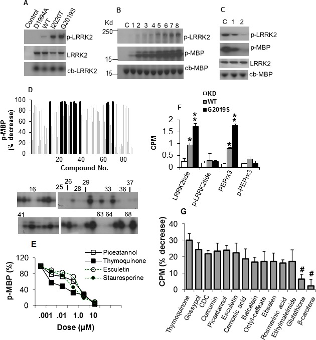
Figure 1.

Antioxidants inhibit LRRK2 kinase activity. (A) Autoradiogram showing levels of p‐LRRK2 in LRRK2 variants (top panel), LRRK2 detected by specific antibody (middle panel) and colloidal blue stain (lower panel). (B) Kinase activity of G2019S on MBP with increasing ATP. Lanes 1–8 contain 0.25, 0.5, 1, 2, 4, 6, 8, 10 μmol/L ATP, respectively. C, no ATP. (C) Inhibition of kinase activity by staurosporine. Lanes 1 and 2 contain 10−3 and 10−1 μmol/L staurosporine. C, no inhibitor. (D) Antioxidants inhibit G2019S activity on MBP. Chart shows percent decrease in MPU of p‐MBP relative to vehicle control (SEM range ± 4.3–12.6, not shown for clarity). Blocked bars represent antioxidants exhibiting reduction at P < 0.01, and correspond to the antioxidant number label on the representative autoradiogram showing the level of p‐MBP. (E) Dose–response curves of G2019S‐mediated MBP phosphorylation as percent of no inhibitor control in the presence of antioxidants. (F) Activity of LRRK2 variants on phosphorylated and nonphosphorylated peptides in the filter‐binding assay, as mean CPM ± SEM (n = 4) of phosphorylated peptide. *,**Significant increase from KD at P < 0.05 and <0.01. (G) G2019S activity on LRRKtide in the presence of antioxidants as percent decrease in CPM ± SEM, corrected from and normalized to control without inhibitor. All values were significant at P < 0.01 except for Glutathione and β–carotene (#). LRRK2, leucine‐rich repeat kinase‐2; p‐LRRK2, phosphorylated LRRK2; MBP, myelin basic protein; MPU, mean pixel unit; p‐MBP, phosphorylated MBP; CPM, counts per minute.
