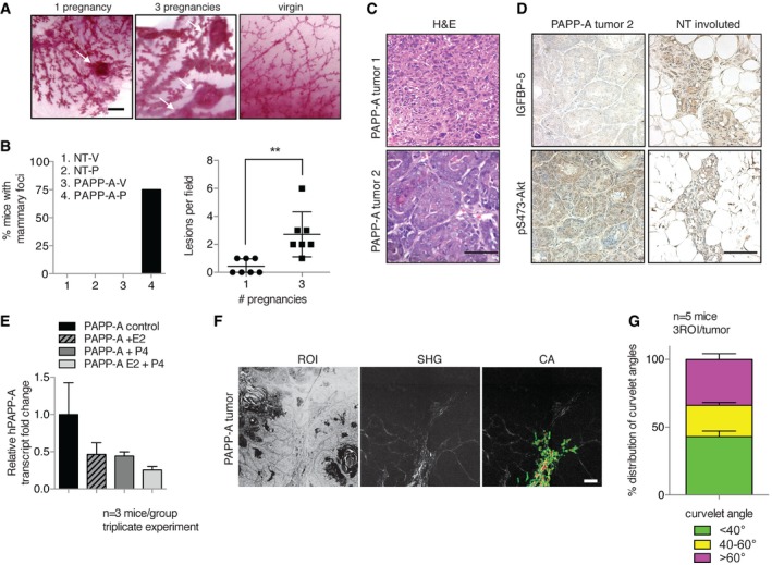
Whole‐mount sections of PAPP‐A mammary glands after 1 or 3 pregnancies and PAPP‐A age‐matched virgin. White arrows indicate lesions. Scale bar: 1 mm.
Left panel: Frequency of mammary lesions in NT virgin (‐V), NT pregnancy (‐P) and PAPP‐A virgin (‐V), PAPP‐A pregnancy (‐P) glands (n = 8 mice except for PAPP‐A‐P: n = 14 mice, seven mice with one pregnancy, seven mice with three pregnancies). Right panel: Number of lesions in PAPP‐A‐P group subdivided into 1 and 3 pregnancies (n = 7 mice, mean ± SEM). Unpaired t‐test (two‐tailed): **P = 0.0038.
Histological sections of two different histologies of a late‐stage (˜14 months) postpartum PAPP‐A mammary tumor. Scale bar: 100 μm.
IGFBP‐5 and pS473‐Akt immunohistochemistry of tumor 2 from (C) and a NT involuted gland, scale bar: 100 μm.
PAPP‐A transgenic mouse mammary glands' PAPP‐A transcript levels in untreated control, treated with estrogen (E2), progesterone (P4), or with both E2 and P4 for 1 week. (n = 3 mice, triplicate experiment, mean ± SD). One‐way ANOVA with Tukey's post hoc test: all P > 0.05.
Imaging of collagen by SHG of PAPP‐A mammary tumor (n = 5 tumors), scale bar: 200 μm.
Analysis of curvelet angle distribution for collagen alignment (n = 5 tumors from five mice, 3ROI analyzed per tumor, mean ± SEM).