-
A
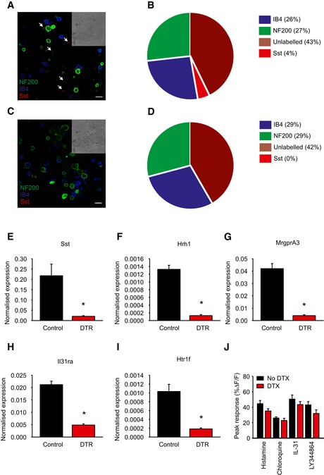
DRG neurons from control AviliDTR/+ mice treated with diphtheria toxin and labeled with antibodies against NF200, Sst, and IB4, and arrows indicate Sst positive. Mice were treated with diphtheria toxin as described below, and DRG cells were isolated, dissociated, and plated on glass coverslips before staining. Scale bar, 30 μm.
-
B
Quantification of the number of NF200, Sst, IB4, and unlabeled neurons in control mice.
-
C
Acutely dissociated DRG neurons from Sst‐Cre::AviliDTR/+ mice treated with diphtheria toxin and labeled with antibodies against NF200 and Sst, and IB4. Scale bar, 30 μm.
-
D
Quantification of the number of NF200, Sst, IB4, and unlabeled neurons in Sst‐Cre::AviliDTR/+ mice after ablation.
-
E–I
Quantitative RT–PCR for the indicated transcripts (Sst, Hrh1, MrgprA3, Il31ra, Htr1f) in DRG from control AviliDTR and Sst‐Cre::AviliDTR mice treated with diphtheria toxin. Values are normalized to ubiquitin levels. Error bars indicate SEM, and asterisk indicates P < 0.05, n = 3.
-
J
Peak calcium flux to the indicated pruritogen in Sst‐Cre::AviliDTR mice treated with vehicle or diphtheria toxin, n = 4–6 mice. Asterisk denotes P < 0.05, t‐test, and error bars indicate SEM.