Figure 4.

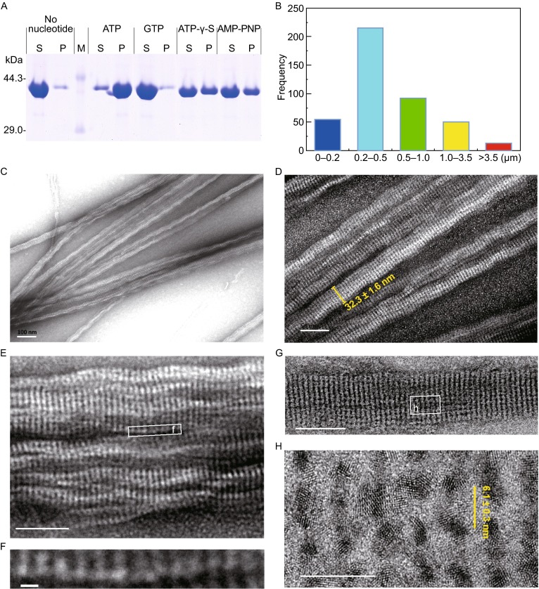
MamK filaments formed in the presence of ATP. (A) MamK polymerization in the presence of various nucleotide states was assessed by a pelleting assay. The supernatant (S) and pellet (P) fractions were analyzed by SDS-PAGE. In the presence of ATP, ATP-γ-S or AMP-PNP, the proportion of the pellet in the total protein was clearly increased compared to the negative control without any nucleotide. (B) The distribution of the filamentous lengths formed in the presence of ATP, Mg2+ and NaCl. The majority of MamK filaments ranged from 0.2 to 0.5 μm in length, and the well-developed filaments were more than 3.5 μm in length. (C–H) Negatively stained MamK polymers visualized by TEM. In vitro, MamK polymerized into well-ordered filaments comprised of multiple filaments in an ATP-dependent manner. Panel C scale bars, 100 nm; panel D scale bar, 50 nm; panel E scale bar, 50 nm; panel F scale bar, 5 nm; panel G scale bar, 50 nm; and panel H scale bar, 10 nm
